Author: 〇ハイデル エムデイヤシン1,2、岩井 治樹1、倉本 恵梨子1、後藤 哲哉1、中村 典史2、山中 淳之1
Affiliation: 1鹿児島大 院医歯 歯科機能形態、2鹿児島大 院医歯 口腔顎顔面外科、3国立科学博物館 人類研究部
Abstract:
The premolar analogy theory says that the premolar tooth is a simplified molar tooth, and that there must be homologies between the premolar and molar cusps. According to this theory, in humans, the buccal and lingual cusps of the upper premolar are believed to be homologous to the mesiobuccal and mesiolingual cusps (the paracone and protocone) of the upper molar, respectively. The purpose of this study is to evaluate the premolar analogy theory from a developmental viewpoint.
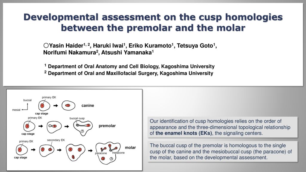
The research design of this study has two fundamental features. Firstly, our identification of cusp homologies relied on orders of appearance and three-dimensional topological relationships of the secondary enamel knots (EKs), the signaling centers which will determine the future cusp positions. We used expression patterns of Shh and Fgf4 mRNAs to identify the EKs. Secondly, we introduced the house shrew, Suncus murinus (Soricidae, Eulipotyphla), as an experimental animal. This insectivorous mammal species possesses full set of tooth types including the canine and premolar, unlike the mouse. We studied cusp formations of the upper canine (C), the upper fourth premolar (P4) and the upper first molar (M1) in this animal.
In the canine development of the house shrew, the primary enamel knot (EK) was identified at the cap stage, and its single structure was kept through the bell stage, which finally formed the single cusp of the canine, suggesting that this could be a basic morphogenetic mode of the unicusped tooth. In the molar development, the primary EK at the cap stage extended distally and split into two domains as the tooth germ grew largely in the distal direction, forming two secondary EKs. The mesial and distal ones formed the paracone and metacone (the mesiobuccal and distobuccal cusps in the human molar), respectively, resulting two buccal cusps in the molar. In the premolar, on the other hand, the primary EK at the cap stage kept its single structure through the later stages, but moved distally, forming the single buccal cusp with sharp distal ridge. These results indicate that the buccal cusp of the premolar is homologous to the single cusp of the canine and the mesiobuccal cusp of the molar. We are going to elucidate the homology of the premolar lingual cusp by exploring the further later stages in the future.
 メールで問い合わせ
コメント